Arteries Diagram / Arterial Supply To The Stomach Complete Anatomy : Spontaneous coronary artery dissection — sometimes referred to as scad — is an uncommon emergency condition that occurs when a tear forms in a blood vessel in the heart.
Arteries Diagram / Arterial Supply To The Stomach Complete Anatomy : Spontaneous coronary artery dissection — sometimes referred to as scad — is an uncommon emergency condition that occurs when a tear forms in a blood vessel in the heart.. The right coronary artery courses in the right atrioventricular groove. Coronary arteries supply oxygenated blood to the heart muscle, and cardiac veins drain away the blood once it has been deoxygenated. We hope this picture human body artery diagram in detail can help you study and research. Arteries are blood vessels that carry blood away from the heart. When the coronary arteries narrow to the point that blood flow to the heart muscle is limited (coronary artery disease), collateral vessels may enlarge and become active.
The narrowed arteries are at higher risk for complete blockage from a sudden. This allows blood to flow around the blocked artery to another artery nearby or to the same artery past the blockage, protecting the heart tissue from injury. Anatomynote.com found human body artery diagram in detail from plenty of anatomical pictures on the internet. We hope this picture common carotid artery diagram can help you study and research. Learn the differences between an artery and a vein.
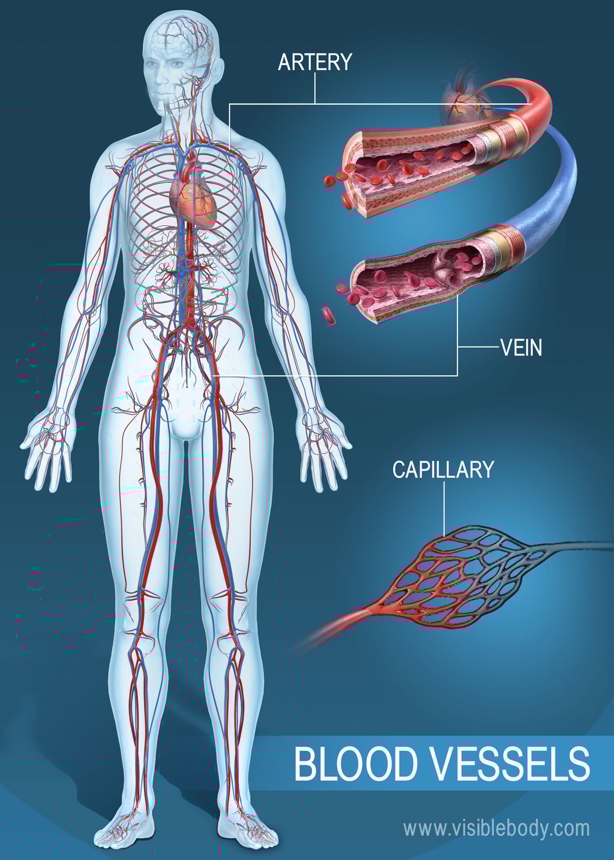
Blood Vessels Circulatory Anatomy from www.visiblebody.com Systemic arteries deliver blood to the rest of the body. Riesige auswahl an cds, vinyl und mp3s. Learn the differences between an artery and a vein. We hope this picture human body artery diagram in detail can help you study and research. Most arteries carry oxygenated blood; Arteries and veins are two of the body's main type of blood vessels. Blood carried by arteries is usually highly oxygenated, having just left the lungs on its way to the body's tissues. Original vintage human anatomy victorian bookplate print 1890s medical diagram veins arteries blood circulatory system of the human body thepapermuseum.
A condition which arises spontaneously or as the result of trauma, where the walls of the artery are split, leading to internal bleeding and disruption of blood flow.
The narrowed arteries are at higher risk for complete blockage from a sudden. In these 101 diagramss, the detailed anatomy of the artery is illustrated in high quality pictures.follow these diagrams to study more about the structures of the artery! Over the years, cholesterol plaques can narrow the arteries supplying blood to the heart. Other sets by this creator. The subclavian artery runs into the axillary region where it becomes known as the axillary artery. Corrective surgery soon after birth is the usual treatment for transposition of the great arteries. Coronary arteries supply oxygenated blood to the heart muscle, and cardiac veins drain away the blood once it has been deoxygenated. Anatomynote.com found common carotid artery diagram from plenty of anatomical pictures on the internet. Anatomynote.com found human body artery diagram in detail from plenty of anatomical pictures on the internet. Arteries of the body diagram. The defect is associated with narrowing of the trachea (windpipe) and bronchi (airways). This bifurcation occurs roughly at the level of the right sternoclavicular joint. The two exceptions are the pulmonary and the umbilical arteries, which carry deoxygenated blood to the organs that oxygenate it (lungs and placenta.
Most arteries carry oxygenated blood; It originates from the heart and branches out into smaller arteries which supply blood to the head region (brachiocephalic artery), the heart itself (coronary arteries), and the lower regions of the body. Over the years, cholesterol plaques can narrow the arteries supplying blood to the heart. The narrowed arteries are at higher risk for complete blockage from a sudden. We shall start at the origin of the carotid arteries.
Blood Vessel Structure And Function Boundless Anatomy And Physiology from textimgs.s3.amazonaws.com Arteries are the blood vessels that carry blood away from the heart, where it branches into even smaller vessels. Arteries are blood vessels that carry blood away from the heart. Scad can slow or block blood flow to the heart, causing a heart attack, abnormalities in heart rhythm or sudden death. Coronary arteries supply oxygenated blood to the heart muscle, and cardiac veins drain away the blood once it has been deoxygenated. A condition which arises spontaneously or as the result of trauma, where the walls of the artery are split, leading to internal bleeding and disruption of blood flow. Coronary arteries supply blood to the heart muscle. Anatomynote.com found human body artery diagram in detail from plenty of anatomical pictures on the internet. The right common carotid artery arises from a bifurcation of the brachiocephalic trunk (the right subclavian artery is the other branch).
The subclavian artery runs into the axillary region where it becomes known as the axillary artery.
We think this is the most useful anatomy picture that you need. Each of these arteries delivers blood to the leg and continues into the foot, with the posterior tibial and fibular arteries forming the plantar arteries and plantar arch that supply blood to the bottom of the foot and toes. Scad can slow or block blood flow to the heart, causing a heart attack, abnormalities in heart rhythm or sudden death. The aorta is the main systemic artery and the largest artery of the body. Over the years, cholesterol plaques can narrow the arteries supplying blood to the heart. Spontaneous coronary artery dissection — sometimes referred to as scad — is an uncommon emergency condition that occurs when a tear forms in a blood vessel in the heart. The right common carotid artery arises from a bifurcation of the brachiocephalic trunk (the right subclavian artery is the other branch). Arteries and veins are two of the body's main type of blood vessels. This is a congenital defect in which the left pulmonary artery branches off the right pulmonary artery, rather than directly from the pulmonary trunk. The defect is associated with narrowing of the trachea (windpipe) and bronchi (airways). Transposition of the great arteries is usually detected either prenatally or within the first hours to weeks of life. A condition which arises spontaneously or as the result of trauma, where the walls of the artery are split, leading to internal bleeding and disruption of blood flow. The coronary arteries wrap around the outside of the heart.
Veins are the blood vessels present throughout the body. The aorta is the main systemic artery and the largest artery of the body. Most arteries carry oxygenated blood; This is a congenital defect in which the left pulmonary artery branches off the right pulmonary artery, rather than directly from the pulmonary trunk. The subclavian artery runs into the axillary region where it becomes known as the axillary artery.
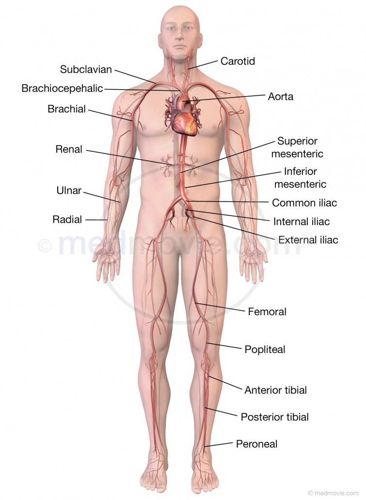
Major Arteries Of The Body Medmovie Com from medmovie.com This is a congenital defect in which the left pulmonary artery branches off the right pulmonary artery, rather than directly from the pulmonary trunk. Though more often occurring with carotid arteries (the other major ones supplying the brain through the neck), vertebral arteries can be impacted. Arteries of the body diagram. Most arteries carry oxygenated blood; These vessels are channels that distribute blood to the body. We think this is the most useful anatomy picture that you need. Coronary arteries supply oxygenated blood to the heart muscle, and cardiac veins drain away the blood once it has been deoxygenated. We hope this picture common carotid artery diagram can help you study and research.
In these 101 diagramss, the detailed anatomy of the artery is illustrated in high quality pictures.follow these diagrams to study more about the structures of the artery!
Though more often occurring with carotid arteries (the other major ones supplying the brain through the neck), vertebral arteries can be impacted. You can see in the artery diagram above the parts of the arteries. Other sets by this creator. Having a baby with transposition of the great arteries can be alarming, but with proper treatment, the outlook is promising. The right coronary artery courses in the right atrioventricular groove. The tunica medica, which is the very muscular middle layer in arteries, is thinner and less muscular in veins. Let's now see how we can revise them with the help of cardiovascular system diagram activities. A condition which arises spontaneously or as the result of trauma, where the walls of the artery are split, leading to internal bleeding and disruption of blood flow. Diagram of the coronary arteries of a human heart poster your walls are a reflection of your personality, so let them speak with your favorite quotes, art, or designs printed on our custom posters! The two exceptions are the pulmonary and the umbilical arteries, which carry deoxygenated blood to the organs that oxygenate it (lungs and placenta. These vessels are channels that distribute blood to the body. Small branches dive into the heart muscle to. The subclavian artery runs into the axillary region where it becomes known as the axillary artery.


0 Komentar